If you are considering ICL surgery, your ophthalmologist may recommend a test called specular microscopy before proceeding. This examination measures the number and condition of endothelial cells on the inner surface of your cornea. Checking the endothelial cell count before ICL is important because these cells help maintain corneal clarity by regulating fluid levels within …


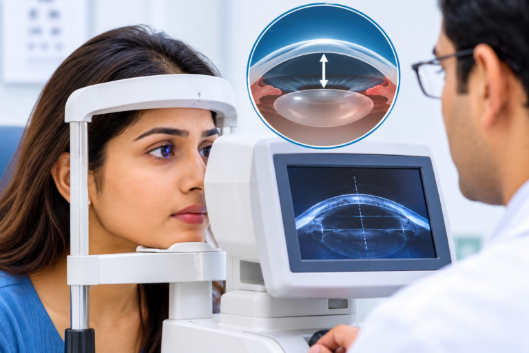
If you are considering ICL surgery, your ophthalmologist may recommend a test called specular microscopy before proceeding. This examination measures the number and condition of endothelial cells on the inner surface of your cornea. Checking the endothelial cell count before ICL is important because these cells help maintain corneal clarity by regulating fluid levels within the cornea. Since an implanted lens sits inside the eye, evaluating the health of these cells helps the surgeon assess surgical suitability and plan appropriate long-term monitoring.
What Are Corneal Endothelial Cells and Why Do They Matter?
The cornea is the transparent front surface of the eye that allows light to enter. Its innermost layer contains a single layer of specialised cells known as corneal endothelial cells.
These cells have an important function: they regulate the movement of fluid from the front chamber of the eye into the cornea.
This keeps the cornea relatively dehydrated and transparent, allowing light to pass through clearly.
Unlike many other cells in the body, corneal endothelial cells have very limited ability to regenerate in adults. When cells are lost, neighbouring cells generally spread out to cover the affected area.
If the endothelial cell population becomes significantly reduced or dysfunctional, the cornea may develop swelling and lose its clarity.
Why Is Endothelial Cell Count Checked Before ICL Surgery?
The endothelial cell count before ICL is an important part of the preoperative assessment because the implanted lens is positioned inside the eye, close to the corneal endothelium.
Although ICL is designed to remain away from the corneal surface, the surgeon needs to evaluate whether the cornea has sufficient endothelial cell reserve.
The examination helps with:
- Assessing corneal health: Determines whether the endothelial cell population is adequate.
- Checking surgical suitability: Helps identify patients who may have an increased risk of corneal complications.
- Planning lens implantation: Supports the assessment of the space between the implanted lens and surrounding structures.
- Establishing a baseline: Provides a reference for comparing endothelial cell counts during future examinations.
- Long-term monitoring: Helps identify changes in endothelial cell density after surgery.
The required cell density depends on factors such as age, anterior chamber depth and the specific ICL model. There is no single universal cell-count threshold suitable for every patient.
How Is an Endothelial Cell Count Test Performed?
The test commonly used to examine corneal endothelial cells is called specular microscopy.
Patients sometimes worry that this examination involves touching the eye. However, specular microscopy is generally a non-contact procedure.
During the examination:
- You sit in front of the diagnostic instrument.
- Your chin and forehead are positioned against the equipment supports.
- You are asked to look at a fixation target.
- The instrument captures images of the corneal endothelial layer using reflected light.
- Specialised software helps analyse the cell density, size and shape.
The examination is generally quick and does not require an incision or injection.
The resulting report provides useful information about the condition of the corneal endothelium.
What Does the Endothelial Cell Count Report Show?
Patients may see several measurements on their specular microscopy report. Understanding the basic terms can make the results easier to discuss with an ophthalmologist.
- Endothelial Cell Density (ECD)
This indicates the number of endothelial cells present per square millimetre (cells/mm²).
A higher cell density generally indicates a greater endothelial cell reserve. However, the result must be interpreted according to the patient’s age and other eye measurements.
- Coefficient of Variation (CV)
This measures variation in the size of endothelial cells.
Greater variation in cell size may indicate that the cells are adapting to cell loss or stress.
- Hexagonality
Healthy endothelial cells commonly have a roughly hexagonal shape. This measurement represents the percentage of cells with six sides.
A reduction in hexagonality may indicate changes in endothelial cell organisation.
- Cell Size
The examination also provides information about the average size of endothelial cells. Changes in cell size may occur as neighbouring cells spread to cover areas where cells have been lost.
These parameters are evaluated together rather than relying on the cell count alone.
What Happens If the Endothelial Cell Count Is Low?
A lower-than-expected endothelial cell count does not automatically mean that a patient has a serious corneal condition. However, it requires careful evaluation before an intraocular lens procedure.
A reduced cell count may be associated with:
- Previous eye surgery or injury.
- Certain corneal conditions.
- Previous inflammation inside the eye.
- Age-related endothelial cell loss.
- Other conditions affecting corneal health.
If the endothelial cell count does not meet the requirements for the proposed ICL model, the surgeon may advise against implantation.
Depending on the patient’s eye condition, alternative vision correction options may be considered. These could include spectacles, contact lenses or suitable corneal laser procedures after a separate eligibility assessment.
Can Endothelial Cells Be Damaged After ICL Surgery?
Endothelial cell loss is an important consideration during and after ICL surgery.
The implanted lens is positioned inside the eye, and factors such as lens positioning, available anterior chamber space and the distance between the implant and corneal endothelium can influence long-term corneal health.
Some endothelial cell loss may occur following intraocular surgery. This is why preoperative measurements and postoperative monitoring are important.
Patients should attend follow-up examinations as advised by their surgeon. Repeat specular microscopy may be recommended to compare the current endothelial cell density with the preoperative baseline.
Regular monitoring helps the ophthalmologist identify significant changes and decide whether additional assessment or intervention is necessary.
What Other Tests Are Required Before ICL Surgery?
An endothelial cell count is only one part of the ICL evaluation. The surgeon may recommend several additional examinations, including:
- Anterior Chamber Depth Measurement: Evaluates the available space between the cornea and natural lens.
- Anterior Chamber Angle Assessment: Examines the drainage angle and helps identify anatomical concerns.
- White-to-White Measurement: Measures the horizontal corneal diameter for lens sizing.
- Corneal Topography: Evaluates the shape and curvature of the cornea.
- Refraction Testing: Measures the patient’s current spectacle power.
- Intraocular Pressure Test: Checks the pressure inside the eye.
- Retinal Examination: Helps identify retinal conditions, particularly in patients with high myopia.
The findings from these examinations help determine whether ICL surgery is appropriate for the individual patient.
Where Can You Get an ICL Evaluation Near Jayanagar?
Patients searching for ICL surgery Jayanagar can consult Kiran Eye Hospital in JP Nagar, Bangalore, for an assessment of their vision and suitability for refractive surgery.
Dr. Kiran Kumar K, Medical Director and Chief Surgeon at Kiran Eye Hospital, holds MBBS, MD (AIIMS, New Delhi), DNB, FAICO (Cornea), FAICO (Refractive Surgery) and MNAMS qualifications.
His professional profile lists expertise in corneal conditions and refractive procedures, including ICL and other vision correction treatments.
During an ICL consultation, patients can discuss their spectacle power, corneal health, endothelial cell count and other diagnostic findings with the ophthalmologist.
For more information, visit the hospital’s ICL/IPCL Surgery service page or Dr. Kiran Kumar’s profile.
Hospital: Kiran Eye Hospital
Location: Ground Floor, Vishnu Towers, 20, ITI Layout, 1st Phase, JP Nagar, Bengaluru, Karnataka 560078.
Frequently Asked Questions
1. Is the endothelial cell count test painful?
No. Specular microscopy is generally a non-contact examination. The instrument captures images of the corneal endothelial cells without touching the eye.
2. What is a normal endothelial cell count before ICL surgery?
Endothelial cell density varies with age and individual eye characteristics. ICL eligibility criteria also depend on the lens model and anterior chamber depth. Your ophthalmologist should interpret the result using the applicable manufacturer’s guidelines.
3. Can endothelial cells grow back after they are damaged?
Adult corneal endothelial cells have very limited regenerative capacity. When cells are lost, neighbouring cells usually spread to cover the affected area rather than replacing the lost cells through significant cell multiplication.
4. Will I need an endothelial cell count test after ICL surgery?
Your ophthalmologist may recommend repeat specular microscopy during postoperative follow-up. The frequency depends on the ICL model, initial cell count and clinical findings.
5. Does a low endothelial cell count mean I cannot undergo ICL surgery?
Not necessarily in every case, but it may make a patient unsuitable for a particular ICL model. The surgeon considers age, endothelial cell density, anterior chamber depth and other eye measurements before making a recommendation.
Book a Consultation
It’s easy and free!